|




|
 Your
editor wrote these articles while a student at an accredited
Chiropractic Medical School. During those days, there were only
two fully Accredited Chiropractic Schools. The ACA gave permission
to reproduce this research at Chiropractic Colleges. It was
through works like this concomitant with far more talented Doctor's of
Chiropractic, which helped the evolution of Chiropractic Medicine; today
Chiropractic Colleges are accredited. Your
editor wrote these articles while a student at an accredited
Chiropractic Medical School. During those days, there were only
two fully Accredited Chiropractic Schools. The ACA gave permission
to reproduce this research at Chiropractic Colleges. It was
through works like this concomitant with far more talented Doctor's of
Chiropractic, which helped the evolution of Chiropractic Medicine; today
Chiropractic Colleges are accredited.
The Theory
of Chiropractic Trauma, which had eluded scientists for centuries
Part 1 Published by the American Chiropractic Association Journal for
the Profession, JACA 1980.
INTRODUCTION
This researcher developed
initial theories, at the University of Minnesota and finalized them
during my first two terms at the Los Angeles College of Chiropractic.
Our then College Dean/neurologist PhD, and I named
the initial theories the "Somato-Neuro-Muscular
and Viscero-Neuro-Muscular Pathway" published by the
American Chiropractic Profession in their official publication the ACA
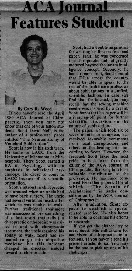
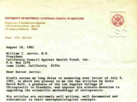
Journal in 1980 (e.g., Then Los Angeles College of
Chiropractic Dean Tran writes, "it is a great pleasure that the college
has given you the spirit to make such contributions to the profession.
And I thank you for the college."). The
Somato-Neuro-Muscular pathway explains a mechanism of
pain and discomfort via direct trauma
to the spine. The Viscero-Neuro-Muscular
Pathway circuits the mechanism of subluxation or altered joint
dynamics via spasm occurring as a direct result of visceral irritation
or disease.
During my third
and forth terms this researcher had written and
subsequently published the mechanism of altered joint dynamics via
"over exertion"
known as the "Physio-Somato-Neuro-Somatic"
reflex systems to finalize my investigations in the
causality of injury and need for manipulative care.
Today, lets review the
Somato-Neuro-Muscular and
Viscero-Neuro-Muscular pathway, which follow:
FORENSIC TEXTBOOK SCIENCE
A subluxation is a
partial dislocation of articulating surfaces of joints, distinct from
luxation, which is a complete dislocation. When a vertebra has been subluxated, ligaments, muscles and other structure in close and delicate
association with the vertebra move.1
Thus,
a point in time arose when direct trauma to the spine had occurred from a
foreign object or a fall on the spine. For example, if the
fifth vertebra thoracic has been subluxated (joint dynamics have been
altered) by trauma or other causes one or more ligaments associated with
it will be stretched! Articular capsules, ligamentum flavum,
supraspinous and intertransverse ligaments are usually the first ones to
develop tension by rotation lateral flexion-rotation of the vertebra.
Interspinous, anterior and posterior longitudinal ligaments also are
involved. If the tension of ligaments reaches a certain threshold,
pressure stimuli will stimulate receptors associated with the ligaments.
From the receptor the stimuli will travel in the spinal nerve to its
cell body located in the dorsal root ganglion and to its axon, to enter
the dorsolateral region of the spinal cord. By following this pathway
the stimuli will ascend in the ipsilateral posterior funiculus of the
spinal cord. As the primary sensory neuron enters the posterior funiculus, it sends a collateral to the posterior
horn.2 This collateral synapses with internuncial neurons associated with the anterior horn,
the lateral horn and the areas of the reticular system in the central
gray area. This system will be discussed with the pain and temperature
pathway.
The primary neuron
which enters the posterior funiculus at this vertebral level (e.g. fifth
spinal nerve) would enter the fascicules cuneatus. The fascicules
cuneatus ascends to terminate in the low medulla oblongata and synapses
with the cell body of the secondary neuron located in the nucleus
cuneatus. From the nucleus cuneatus, the secondary neuron decussates to
form the internal arcuate fiber into the posterior portion of the medial
lemniscus. This neuron now ascends through the pons as part of the
medial lemniscus. In the pons, the medial lemniscus rotates so that its
posterior portion becomes medial and its anterior portion becomes
lateral. The secondary neuron is located in the medial portion of the
medial lemniscus. From there, it ascends to terminate in the
posteriolateralventral nucleus of the thalamus. The
posteriolateralventral nucleus of the thalamus represents the site of
the cell body of all tertiary neurons of this pathway. From there, the
tertiary neuron ascends through the posterior limb of the internal
capsule, through the corona radiata to end in the post central gyrus,
also know as the somesthetic cortex, area 3,1,2.
At this level the brain would acknowledge
the tension of ligaments involved.3
The primary neuron
also synapses with association (internuncial) neurons in the posterior
horn. These internuncial neurons will synapse with the reticular neurons
located in the reticular core of the central gray area. 4 From there, the
spinothalmo-reticulo-limbic pathway ascends to higher centers including
the hypothalamus. Impulses from the hypothalamus will descend through
reticulospinal pathways to the lateral horn and
at the anterior horn at the
level of the spine. From the lateral horn of appropriate levels stimuli
will be carried to the respective organs by way of sympathetic fibers.
This preganglionic neuron enters the sympathetic chain ganglia to
synapse with a postganglionic neuron. From there, the postganglionic
neuron goes to smooth muscles of bloods vessels.5
This may cause vasoconstriction.
The reticulospinal
fibers, which terminate into the anterior horn, will synapse with
lower
motor neurons. The lower motor neuron goes to striated muscle in the
area of the altered joint dynamics
(Chiropractic Subluxation) on the ipsilateral side. This innervation increases the tonus of the associated
back muscles.6 The muscles increase their energy output
which causes an increased contraction of arterial
blood vessels, engaging endothelium triggers which
release endothelan-1 and prostanoids further causing vascular
constrictions, ischemia,
increased lactic acid, bradykininn, P factor, hydrogen ions, potassium
ions, and metabolites formed by the
breakdown of muscle (waste materials),
are all produced.7
They subsequently
accumulate in
the tissue spaces, with resulting
increased interstitial fluid colloid osmotic pressure, increased muscle
fluid in the interstitial space, increased muscle stiffness,
over-stimulation of nerve endings by the presence of
trapped metabolites, temporary local soft tissue acidosis
resulting from lactic acid
and the hydrogen ions,
(Please see Part 2. Subluxation caused by
muscular overexertion sprain/strain) pain, muscular spasticity with the resultant altered joint dynamics
(again restricted, strained and
sprained spinal joints).
From the
stretching of the ligaments associated with the subluxation, Golgi end
organs connected with the conscious proprioceptive pathway conduct their
impulses to the dorsal root ganglion to enter the central nervous system
at the Lissauer’s marginal zone and synapse with the nucleus proprius.
From there, the tertiary neuron ascends to the post central gyrus areas
3, 1, 2 of the somesthetic cortex. This tract follows closely the path
of the pain and temperature pathway (internal capsule to corona radiata to
somesthetic cortex).8
When the primary
neuron synapses with the secondary neuron of the pain and temperature
pathways at the spinal level, intersegmental neurons generally carry the
impulses from one to two levels above and below the spinal level of
origin. 9
With several levels
stimulated, muscles spasticity may spread to levels above and below the
area of altered joint dynamics (Chiropractic Restriction-Subluxation).
This condition could lead to subluxations at other levels because
increased muscle tonus produces areas of altered joint dynamics (The
Chiropractic Subluxation Complex). This cyclic
pathway, which has been presented and has stood the test of Judicial
Review, Justice Department Review, Scientific Review, and Chiropractic
Review is known as the "Somato-Neuro-Muscular Pathway".
Another common
type of back pain manifested by altered joint dynamics (The Chiropractic
Subluxation) stems from internal organ irritation. For example, if too
much hydrochloric acid has been secreted within the stomach the
irritation may stimulate the submocosal sensory fibers of the Meissner’s
plexus .10
These pain fibers enter the central neurons system and from
the lateral spinothalamic tract, end in the cortex, as previously
described. Because the primary neuron of the pain pathway synapses in
the posterior horn, it is associated with a circuit connecting
internuncial neurons to the lateral horn, anterior horn and to the
reticular system.
Muscles spasticity is produced via the anterior horn connections.
Synapses from the lateral horn cause vasoconstriction of blood vessels
by the sympathetic system leading to ischemia, increased lactic acid,
increased P factor, Hydrogen and Potassium ions, thus an increase in
pain. The muscles’ spasticity can cause altered joint dynamics in the
back and a vicious cycle results.
This visceral
pathway, which has stood the test of Judicial Review, Justice Department
Review, Scientific Review, and Chiropractic review is called the
"Viscero-Neuro-Muscular
Pathway". Today, we have firmly established that
the pancreas can refer pain to the
lumbar spine, the Gall Bladder refer to the tips of the scapula, the
heart to the Jaw, left shoulder arm and so forth.
DISCUSSION:
Although
Chiropractic Medicine has repeatedly proved itself in practice, this
proof should be communicated to the medical world in its own language; a
task that can only be accomplished by demonstrating chiropractic
principles first in theory, then by clinical trials and by demonstrating
said principals in the laboratory. Thus, through work such as this and
laboratory experiments the medical profession and the chiropractic
profession will, one day, work together to make the
world a healthier
place in which to live.
Look to the
"Somato-Neuro-Muscular
Pathway" and the "Viscero-Neuro-Muscular Pathway" as the first bridge to
new and greater shared communications with our partners in health care,
the medical community. "Health
Care is a team effort and should not be divided"
(Neff 1980 The Neuroanatomy of Vertebral Subluxations-ACA)".
“Ye
shall know the truth, and the truth shall make you free.
”John, viii, 32
REFERENCES:
1.
Gray's Anatomy, 35th British edition, W.B.
Saunders Company, 1978, pp 413-414.
2.
Carpenter: Human Neuroanatomy, 7th edition, Williams and Williams
Company 1977, pp 238-242. Fig 10-1
3.
(i) Gray's Anatomy 35th British edition, W.B. Saunders Company 1978, pp
824-825. (ii) Carpenter: Human Neuroanatomy, 7th edition, Williams and
Williams Company, 1977, pp 238-242.
4.
Gray's Anatomy, 35th British edition, W.B. Saunders Company 1978, pp
836-837, 888-890.
5.
Guyton: 5th edition, W.B. Saunders Company 1976, pp 768-773, 776-778.
6.
Carpenter: Human Neuroanatomy, 7th edition, Williams and Williams
Company, pp 255-261.
7.
(i) Lewis, T.; Pain, The MacMillan Co., 1943, pp 99-103. (ii) Kor, I.M.:
The Neurobiologic Mechanisms in Manipulative Therapy, Plenum Press,
1978, p.457. (iii) Buergen and Tobis; Approaches to the Validation
of Manipulative Therapy, Charles C. Thomas, 1977, p. 191.
8.
Carpenter: Human Neuroanatomy, 7th edition, Williams and Williams
Company, 1977, pp 245-248.
9.
Guyton: 5th edition, W.B. Saunders Company, 1976, p. 652.
10.
Guyton: 5th edition, W.B. Saunders Company, 1976, p. 852
EDITORS NOTE:
Looking back, I have noticed
a very interesting advertisement on the back side of the facing page of
my article within then official publication for the Chiropractic
Profession, the Journal of the American Chiropractic Association.
The add is from the "International Academy of Trial Lawyers" for
featured speaker, F. Lee Bailey. Under his picture it is written,
Internationally known attorney, author, teacher and lecturer, on
"Successful Management of Personal Injury Litigation...."
You just cannot make this stuff up.
by
Scott D. Neff, DC CFE DABFE FFABS FFAAJTS
SCIENTIFIC INVESTIGATION INTO SUBLUXATION STRAIN
SPRAIN PART TWO
BY SCOTT D NEFF, DC DABCO MSOM MPS-BT CFE DABFE FFABS
FFAAJTS

|
![]()