VAGINAL WET PREPS
A female patient presents with itching, odor, discharge or any combination of therein. This patient is
immediately referred out to your consulting Gynecologist. During examination the vaginal ph, and discharge is noted and a sample is collected. The physician can either send this to the lab or go into their own lab. Of course the lab tech wears their gloves and lab coat. Aside the microscope would be placed their plastic-backed towel, rack, and lab tray/basket. The vaginal sample is placed with 1/2 millimeter of saline into a test tube with a cotton swab and toped. The top is temporarily removed, and with a bulb pipette, gently but thoroughly mix the material on the swab with the saline until the suspension is homogenous. With the bulb pipette place one drop of the suspension on a slide and the cover slip is placed over it properly to avoid trapping air bubbles. Do not use a free-falling drop. The drop size should be approximately 1/3 the size of the cover slip. Apply the cover slip by placing one corner of the cover slip on the edge of the visual slide area and releasing the rest of the cover slip to drop quickly on the slide to cover the center visualization area. One drop of the vaginal sample is placed on another slide with 1 drop of 10% KOH solution. Immediately waft with the hand while sniffing for a fishy or amine odor and then quickly place the cover slip on avoiding any air bubbles. The fishy odor indicates trichomonias or bacterial vaginosis as KOH volatilize the Amines. Examination would be relative to the following: a. Squamous Epithelial cells b. PMN's or Poly's c. Clue Cells which are Squamous epithelial cells coated with enough coccobacillary bacteria that 75% of the boarder is obliterated with bacteria. These cells indicative of bacterial vaginosis look like epithelial cells with glue on them looking like the cell is pressed in sand. d. Yeast cells such as Pseudohyphae or long tubular branching cells or Yeast buds appear like a shoe print (heal and soul). In this case usually the Doderlein's bacillus, a large gram-positive microorganism commonly found in the vagina also known as vaginal protective flora has been disrupted. e. Red blood cells SALINE PREP: On the saline prep one may find Epithelial cells, Trichomonads, PMN's and Clue Cells. If the Yeast cells are not superimposed by other cells they can be visualized as well as sperm and bacterial cells. The cells must be examined quickly as Trichomonads lose their moving abilities within 15-20 minutes and then become undistinguishable from other cells. KOH PREP: On the KOP prep one may only find Yeast cells and all other cells will be lysed. MICROSCOPY:
The lab technician cleans the microscope lens with lens paper and solution as well as cleaning again when your done. They place the Saline slide sample on the microscope stage. Rotate the 10x objective into place. Turn the microscope light on. Use the course adjusting knob to bring the objects into focus. Adjust the condenser or diaphragm for optimal contrast if necessary. Next they rotate the 40x objective into place. Raise the light level and adjust the fine tuning knob now to bring the sample into focus. Re-adjust the condenser or diaphragm if necessary. Begin reading. If they have difficulty visualizing the saline sample re-use the 10x for the wide view to find pseudohyphae or trichomonads and then switch back to the 40x for confirmation. They use the z-pattern for your eyes visual analysis or the channel method prior to notation. They note the Hi power field quadrant for WBC, RBC, Sperm and clue cells. Quantitate these elements and report each category as the number/hpf (e.g., 5-10 WBC/hpf.) which is done by multiplying the 1 quadrant findings by four and writing the number of cells found. They note the Semi-quadrant counts for Bacteria, Yeast, their shape or if trichomonads are found. Semiquantitate these elements and report using trace 1+, 2+, 3+ or 4+ system of denoting concentration/density of the materials. If not seen, note not seen. TRICHOMONAS VAGINITIS Saline Motile trichomonads KOH negative WHIFF positive Ph > 4.5 BACTERIAL VAGINOSIS Saline Clue cells-one in each of 10 fields or 1 out of 5 epithelial cells should be clue cells. KOH negative WHIFF positive Ph > 4.5 YEAST VAGINITIS Saline Budding yeast and/or pseudohyphae KOH Budding yeast and/or pseudohyphae WHIFF negative Ph <4.5 by
Dr. Scott D. Neff, DC DABCO MSOM MPS-BT CFE DABFE
FFABS FFAAJTS,
graduate
of the University of Health Science Antigua School of Medicine. 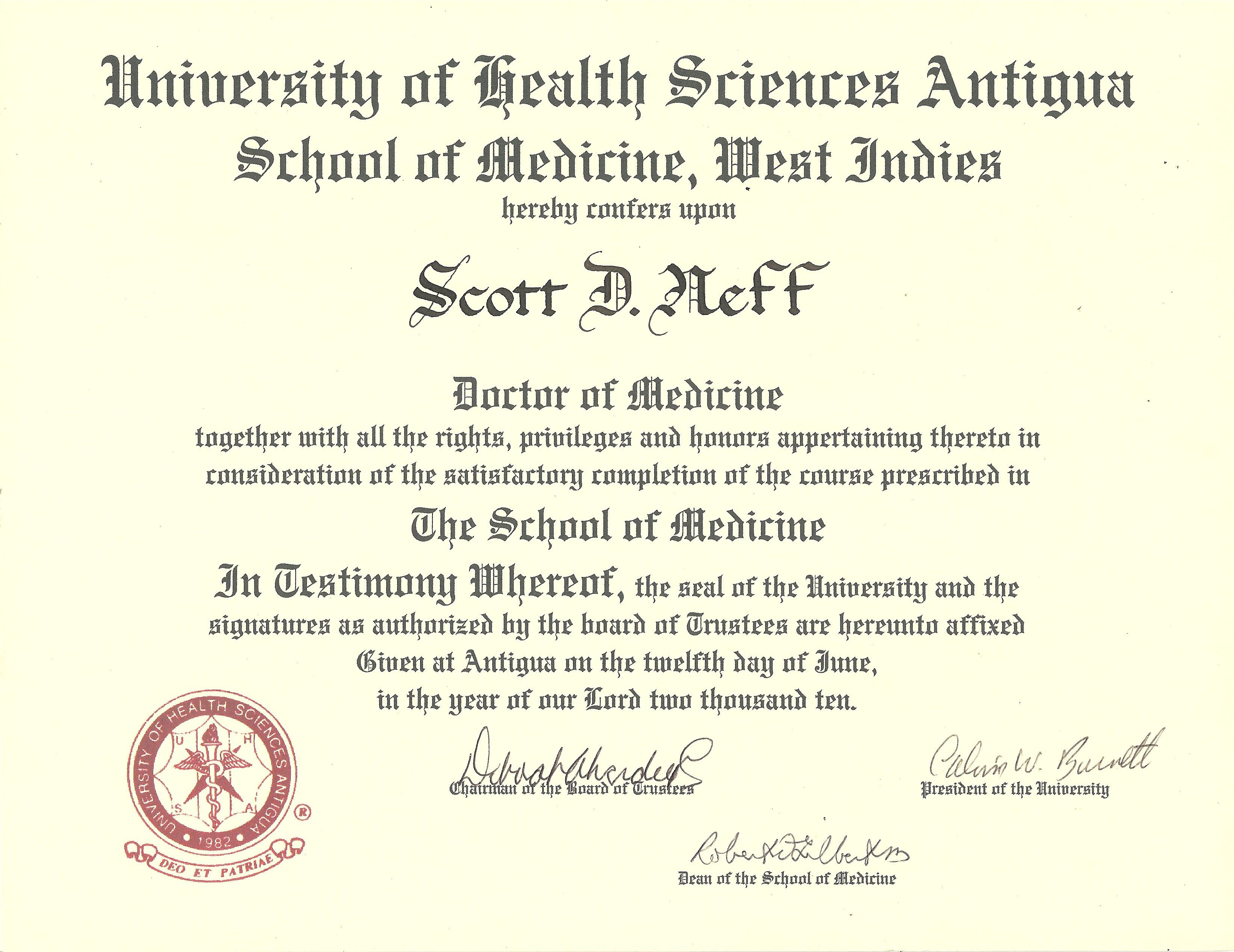
|